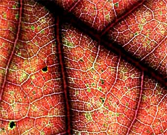
There's a lot more to leaf veins that just noticing the pretty patterns they form, such as those in the leaf of an autumnal Black Oak, Quercus velutina, shown at the right. In that picture, the big, blackish line across the top, right, is the leaf's midvein originating at the leaf's stalk, or petiole, with secondary veins branching from the midvein.
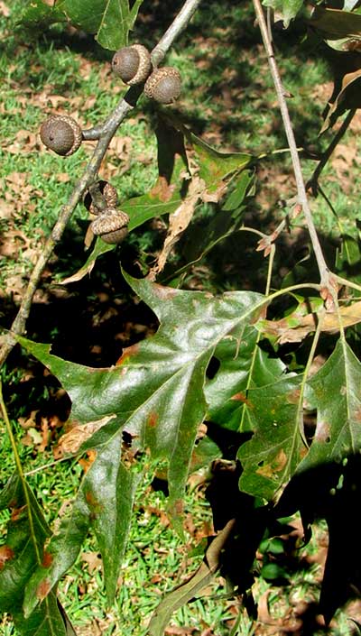
At the left, on a Black Oak tree, you can see how the leaf's petiole continues up through the leaf, becoming the midvein. In the above close-up picture, notice that secondary veins have smaller veins branching from them, then those smaller veins branch again. In the end, that amounts to a lot of veins. It's been estimated that if the veins in a single elm leaf were placed end to end they would be more than 700 feet long (210 m).
In a leaf, the veins' branching and rebranching is like the system of roads leading from a big city. When you leave a city on a main road, then continually keep turning onto smaller and smaller off-roads, eventually you come to a dead end. In leaves, eventually you end up at a more or less random spot in the leaf. With both city roads and leaf veins, the idea is to transport traffic to and from final destinations. Roads carry people and the things people need and produce. Leaf veins carry water and, dissolved in the water, things the plant needs and produces.
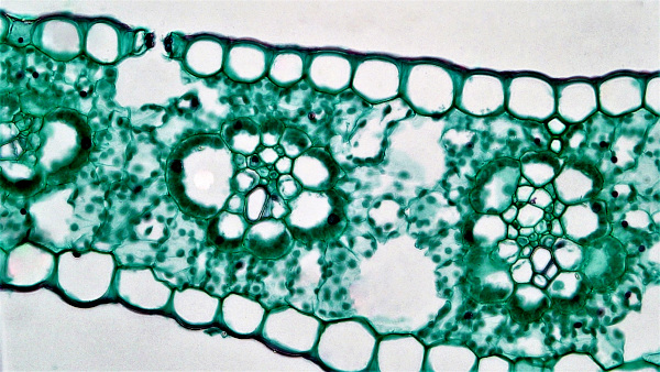 Cross section of corn leaf, Zea mays; courtesy of Berkshire Community College Bioscience Image Library in Massachusetts, and Wikimedia Commons
In the picture at the right showing a microscopic cross section of a leaf of corn, Zea mays, you can see the leaf's individual cells. Large cells covering the leaf's upper and lower surfaces form the skin-like epidermis. Most of the leaf's tissue consists of small, irregularly shaped, closely packed mesophyll cells inside which photosynthesis takes place. The tiny, dark specks inside each mesophyll cell are chlorophyll-containing, photosynthesizing chloroplasts. The two dark-fringed clusters of different-sized cells surrounded by mesophyll cells are vascular bundles.
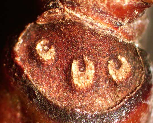
Within each vascular bundle, the cells nearest the leaf's upper surface are xylemcells, while the bottom-facing ones are phloem cells. Xylem cells conduct water and dissolved nutrients upward from the root, while phloem cells convey sugars and other food-to-energy (metabolic) products downward from the leaf's photosynthesizing cells. In the leaf scar shown at the left, resulting from the leaf of a Sweetgum tree, Liquidambar styraciflua, naturally breaking from a stem in the fall, the three horseshoe-shaped items are bundle scars. bundle scars form where veins containing xylem and phloem cells break between the petiole and the stem.
LEAF VEINS DISPLAY MANY PATTERNS
Atop this page we saw the branching and rebranching reticulate venation of a dazzlingly red leaf of a Black Oak. At the right, the opposite extreme of such free-form venation appears in the leaf of a Devil's Shoestring, Nolina lindheimeriana, a shrubby, evergreen perennial of limestone hills, open woodlands and scrubland of southwestern Texas. It shows the highly regimented parallel venation typical of monocots. This monocot belongs to the Agave Family.
At the left, veins in the leaf of a Burhead, Echinodorus subalatus, the main or primary veins are considered to be parallel, but the secondary veins are said to be reticulate. However, the primary veins are only approximately parallel, for they all originate at one point at the leaf's base, plus the secondary veins themselves are somewhat parallel. In other words, Nature makes no effort to fit all Her creations into humanity's neat categories, though the categories still are useful if we want to talk and think about things.
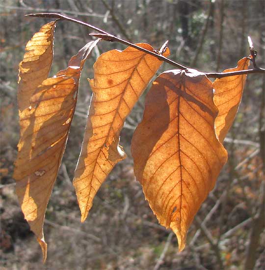
One of the most commonly seen leaf venation patterns is shown at the right, on wintertime-persisting leaves of the American Beech, Fagus grandifolia. With strong, regularly spaced secondary veins so neatly branching off the central midrib, this is pinnate venation.
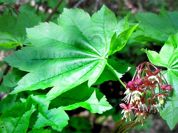
At the left, a leaf of the Vine Maple, Acer circinatum, shows palmate venation, with its primary veins radiating from one point at the leaf's base.
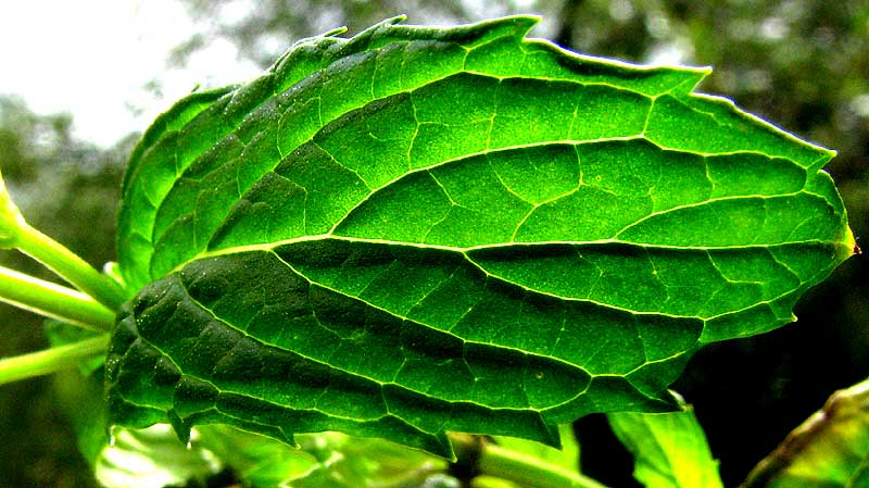
At the right, the leaf of a garden Spearmint, Mentha spicata, shows two noteworthy leaf-vein features. First, the secondary veins in many leaves are fairly straight, and often end at a margin tooth or pointed lobe. The Spearmint leaf's secondary veins, however, curve toward the leaf tip and end within the blade. Such leaves show arcuate venation. Also, notice that major veins are sunken below the level of most of the blade surface. Such veins are said to be impressed.
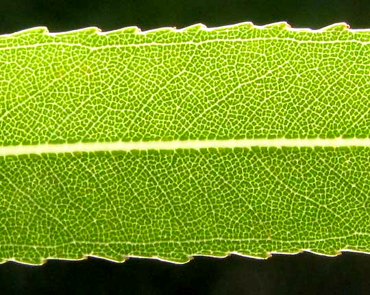
At the left, the leaf of the Black Willow, Salix nigra, shows a feature that's very helpful during the identification process, for only certain groups of plants have leaves doing what's shown. Notice that a vein runs along each leaf margin a small distance inside the leaf. These veins connect with the tips of secondary veins. Leaves with such veins are said to have marginal leaf veins.
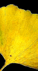
At the right, a leaf of the Ginkgo tree, Ginkgo biloba, displays dichotomous venation. Note that each vein splits in two, then those veins split, on and on, more or less staying parallel to one another and never reticulating.
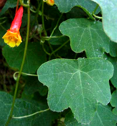
Sometimes petioles attach to their leaf blades not at the very base, as with the vast majority of leaves, but within the leaf's margin -- the way an umbrella's handle attaches to its canopy. Such is the case with the leaf at the left, of a wild nasturtium species, Tropaeolum emarginatum. It displays peltate venation. Notice that its primary veins radiate from a point within the blade's surface, not from at its margin at the leaf base.
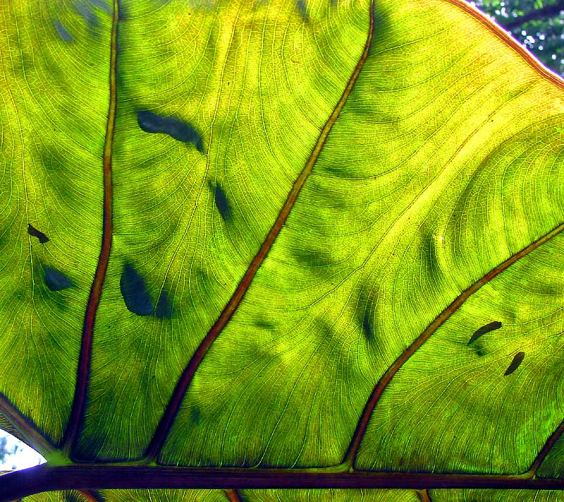
Finally, sometimes it just seems impossible to precisely classify certain leaf venations. Behold what the veins do on the tropical Giant Taro, Alocasia macrorrhiza: